What does a neuron look like?
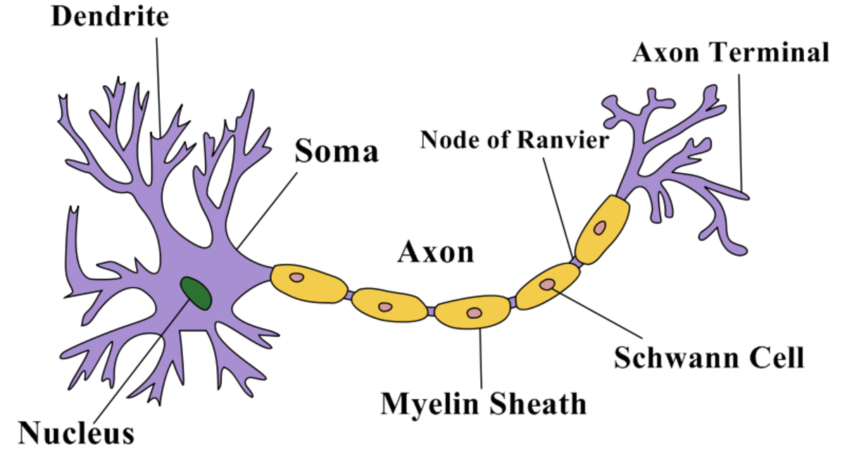
neuron, basic cell of the nervous system in vertebrates and most invertebrates from the level of the cnidarians (e.g., corals, jellyfish) upward.A typical neuron has a cell body containing a nucleus and two or more long fibres. Impulses are carried along one or more of these fibres, called dendrites, to the cell body; in higher nervous systems, only one fibre, the axon, carries the impulse.
Neurons
There are three main types of neurons: Motor neurons make the connection between the brain and muscles throughout the body. These neurons transmit electrical impulses containing information to skeletal muscles and smooth muscles. Motor neurons control all of our body movement. Sensory neurons are neurons that let us feel sensation.
Histology of the Nervous System (The Neuron) Part 1
An Easy Guide to Neuron Anatomy with Diagrams By Olivia Guy-Evans, MSc Updated on November 9, 2023 Reviewed by Saul Mcleod, PhD Neurons are the information processing units of the brain responsible for sending, receiving, and transmitting electrochemical signals throughout the body.

FileNeuron1.jpg Simple English Wikipedia, the free encyclopedia
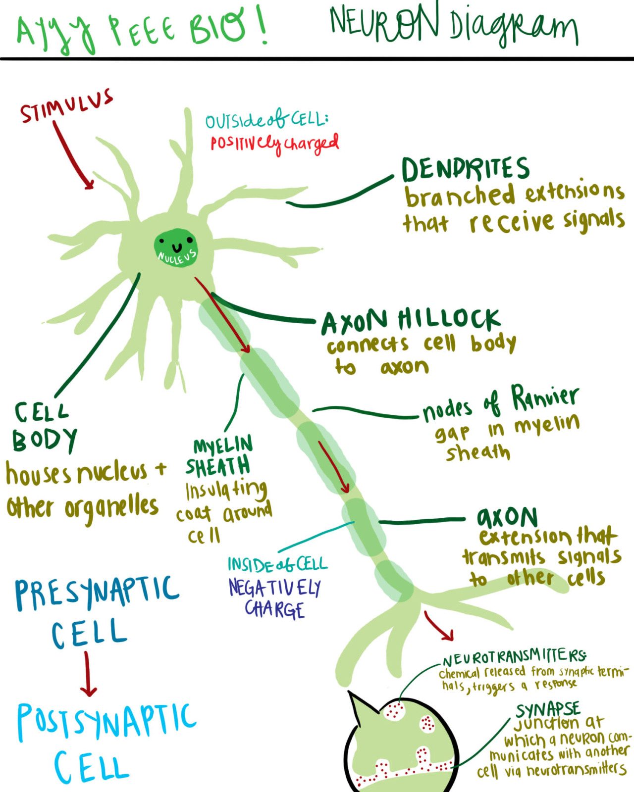
The parts of the neuron have been labeled. Your challenge is to write the correct name for each part and explain what it does. If you need some help, visit the web article listed below. Neuron Anatomy Activity Synapses: Send electrical impulses to neighboring neurons.
Structure and types of neuron (The nervous tissue) Online Science Notes
Nervous system Schematic of an anatomically accurate single pyramidal neuron, the primary excitatory neuron of the cerebral cortex, with a synaptic connection from an incoming axon onto a dendritic spine Neurons are the primary components of the nervous system, along with the glial cells that give them structural and metabolic support. [4]
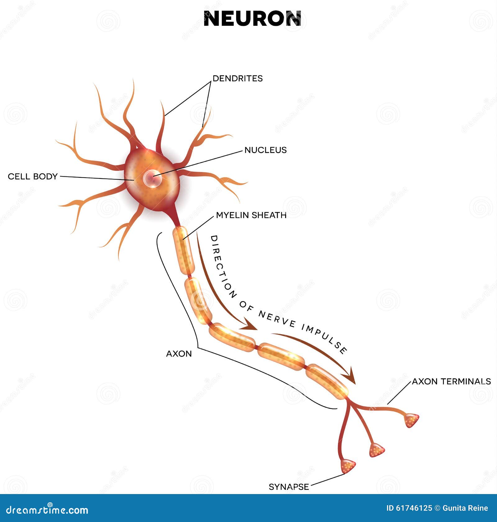
Labeled Diagram of the Neuron Stock Vector Illustration of anatomical, peripheral 61746125
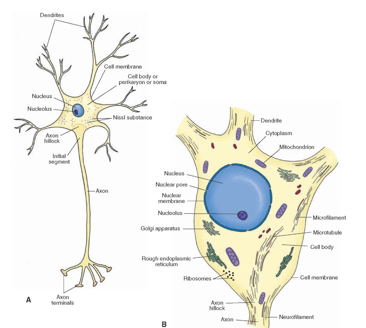
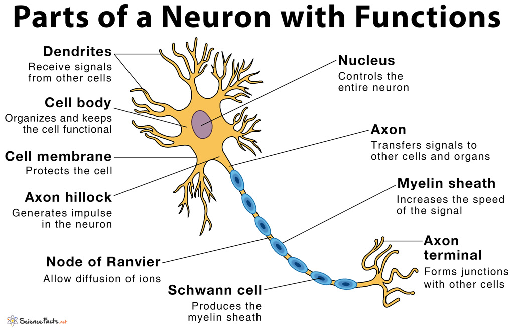
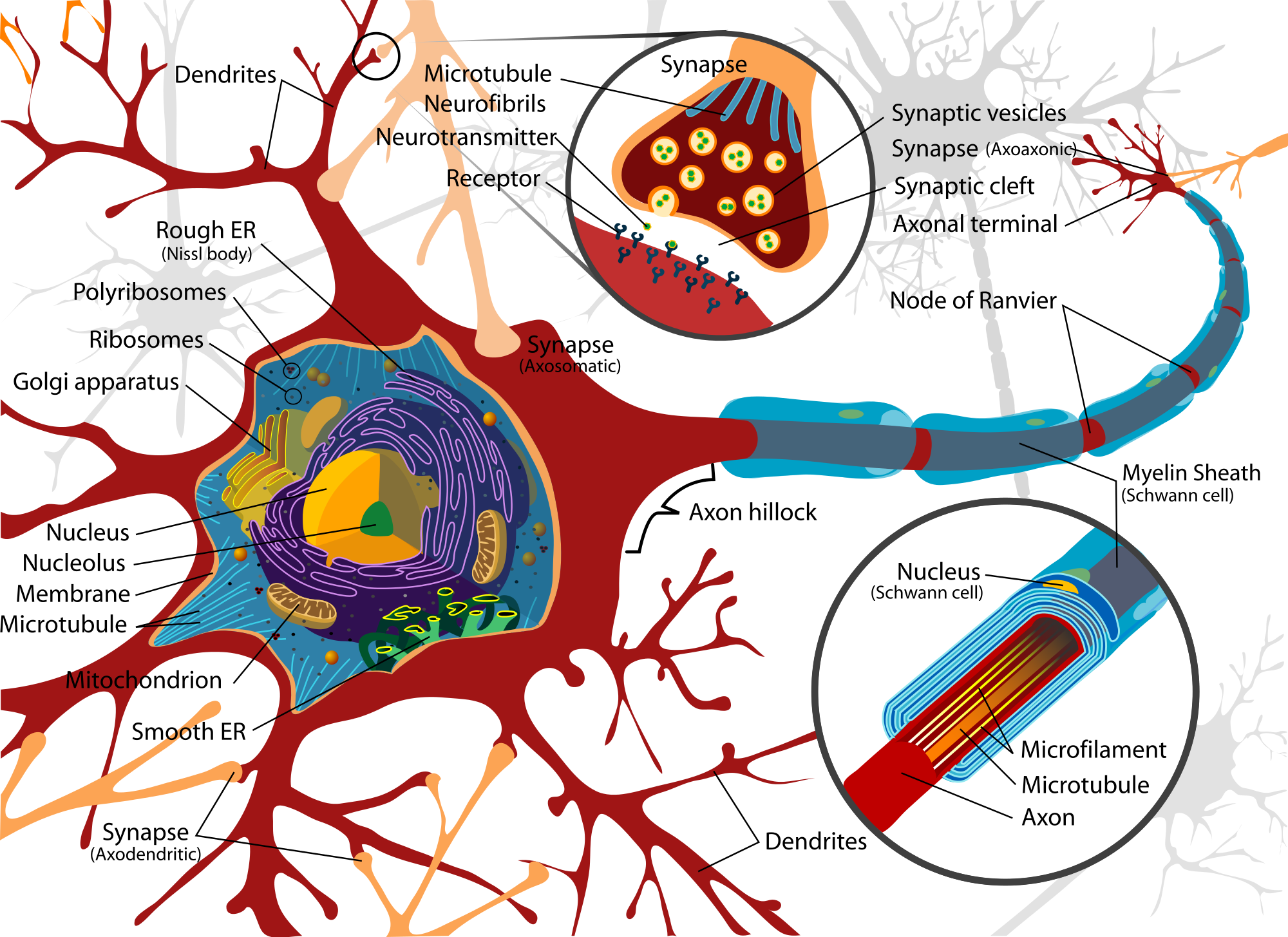
The cell body of a neuron, also known as the soma, is typically located at the center of the dendritic tree in multipolar neurons.It is spherical or polygonal in shape and relatively small, making up one-tenth of the total cell volume.. The functionality of the neuron is highly dependent on its cell body as it houses the nucleus, which contains the genetic material (DNA) of the cell as well as.

Health Lab 4.1 Introduction
Classes of neurons Based on their roles, the neurons found in the human nervous system can be divided into three classes: sensory neurons, motor neurons, and interneurons. Sensory neurons Sensory neurons get information about what's going on inside and outside of the body and bring that information into the CNS so it can be processed.
A diagram of a neuron and its functions. a study in chartreuse
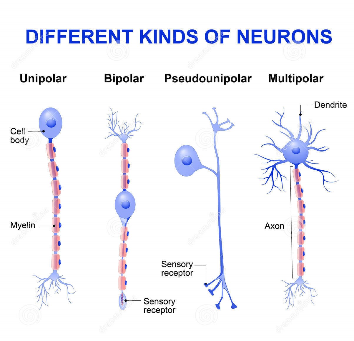
Describe the basic structure of a neuron Identify the different types of neurons on the basis of polarity List the glial cells of the CNS and describe their function List the glial cells of the PNS and describe their function Nervous tissue is composed of two types of cells, neurons and glial cells.
Structure of a Neuron Owlcation
Cerebellum - molecular, Purkinje, granular layers. Peripheral nerves - epineurium, perineurium, endoneurium. This article will explain the histology of neurons, providing you with information about their structure, types, and clinical relevance. It will also cover briefly the histological layers of the central and peripheral nervous systems.

Anatomy Of A Neuron ANATOMY
At the synapse, the firing of an action potential in one neuron—the presynaptic, or sending, neuron—causes the transmission of a signal to another neuron—the postsynaptic, or receiving, neuron—making the postsynaptic neuron either more or less likely to fire its own action potential. Schematic of synaptic transmission.
Neuron Study Guide Inspirit Learning Inc
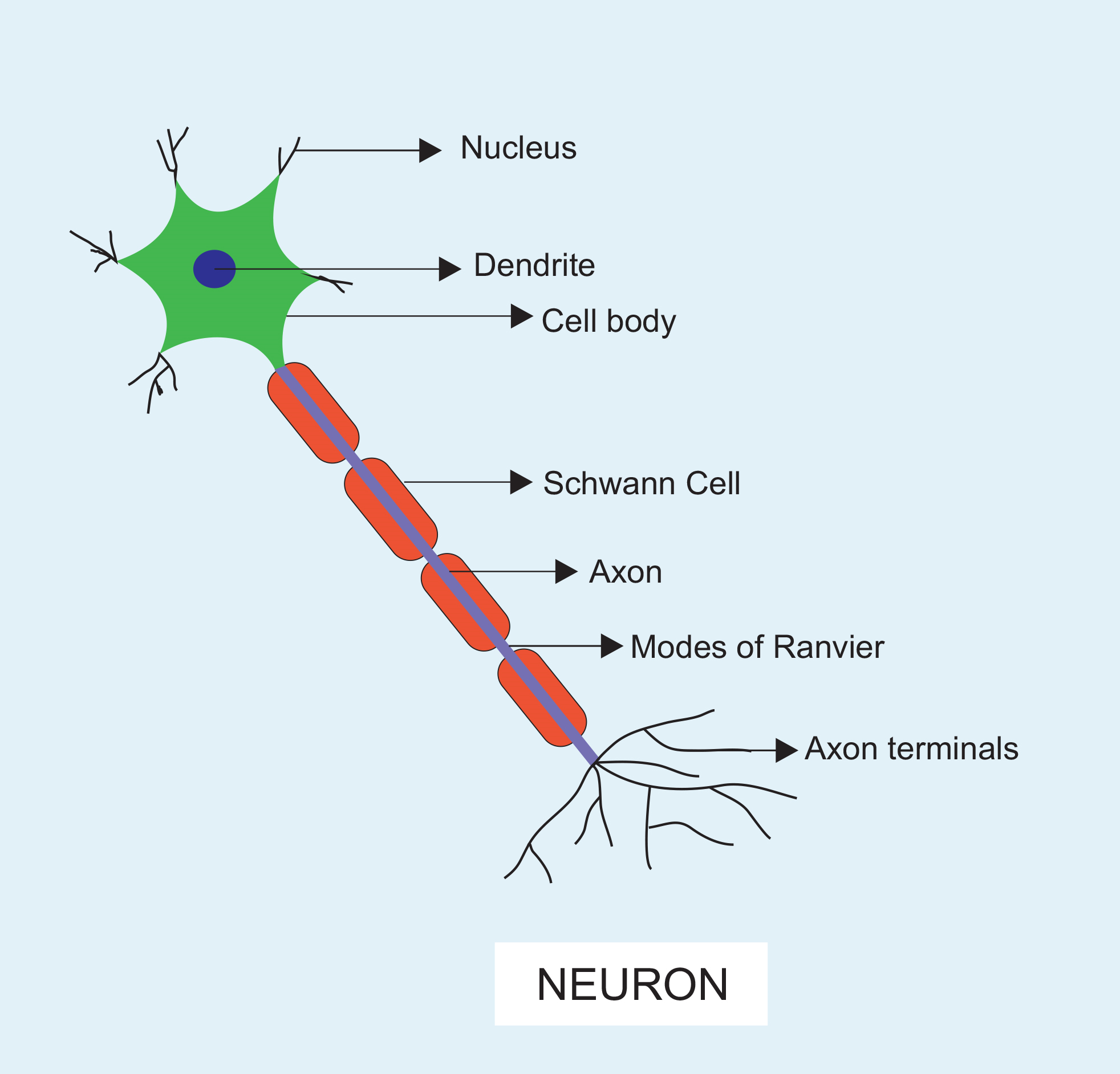
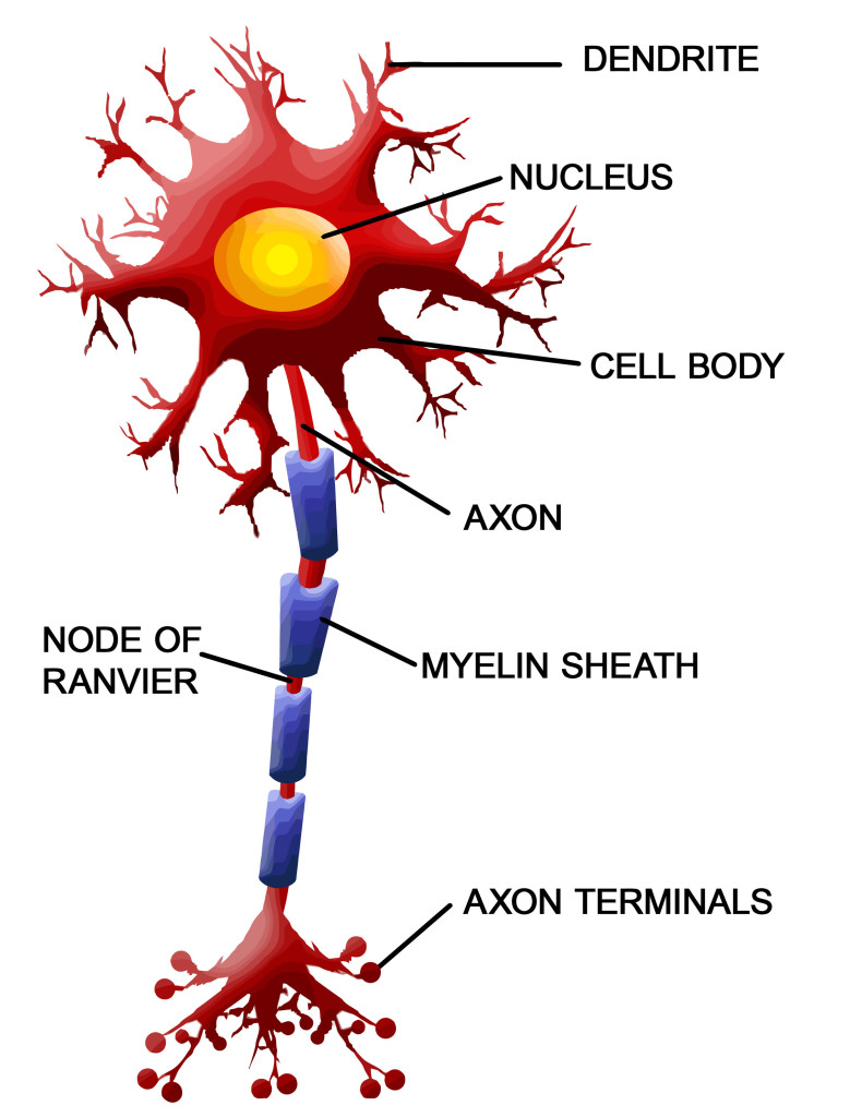
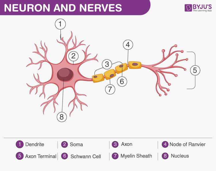
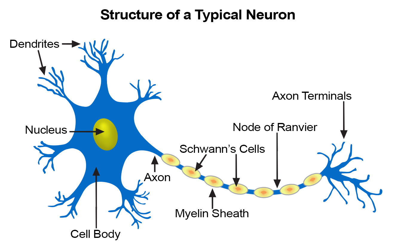
A diagram of a neuron also known as the nerve cell is useful as a visual tool to illustrate the various components of the neuron. It also helps us to understand the functions of the neuron.. Labeled Diagram of a Neuron. The different components of the neuron are illustrated below, with each area specified and labeled. Structure of Neuron.
The Neuron External Structure and Classification Interactive Biology, with Leslie Samuel
A Labelled Diagram Of Neuron with Detailed Explanations Biology Biology Article Diagram Of Neuron Diagram Of Neuron A neuron is a specialized cell, primarily involved in transmitting information through electrical and chemical signals. They are found in the brain, spinal cord and the peripheral nerves. A neuron is also known as the nerve cell.
What does a neuron look like? Biology Questions
The nervous system is a network of neurons whose main feature is to generate, modulate and transmit information between all the different parts of the human body. This property enables many important functions of the nervous system, such as regulation of vital body functions ( heartbeat, breathing, digestion), sensation and body movements.
Nerve Cell Diagram Labeled ClipArt Best
A detailed diagram of a neuron, showing the synapse, cell body, axon, and dendrites. To unlock this lesson you must be a Study.com Member.
Nerve Tissue SEER Training
Parts of a Neuron Diagram Although they have a characteristic elongated shape, they vary widely in size and properties based on their location and type of functions they perform. While they have the common features of a typical cell, they are structurally and functionally unique from other cells in many ways.

Neuron Diagram Straight from a Scientist
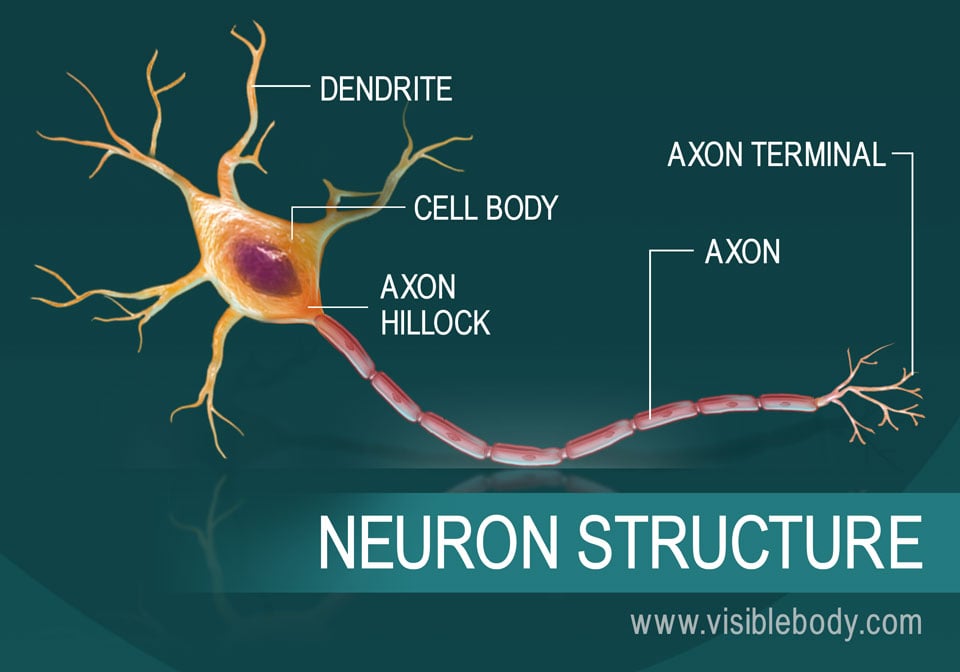
About Transcript Neurons (or nerve cells) are specialized cells that transmit and receive electrical signals in the body. Neurons are composed of three main parts: dendrites, a cell body, and an axon.
